Restoring mitochondrial and lysosomal function in neurodegeneration
Reprogramming cells. Repairing neurons. Restoring lives.
Endurance Bio develops first-in-class oral small molecules designed to restore mitochondrial and lysosomal function through the PGC-1α–TFEB axis. Our lead programs, T-168 and T-621, target interconnected cellular systems that become dysfunctional in neurodegenerative, metabolic, muscle and age-related diseases - rather than a single downstream pathway. Our lead clinical program is Parkinson's disease.
- T-168 and T-621 restore mitochondrial and lysosomal function in dopaminergic neurons, and restore dysfunctional and inflamed microglia to their normal, homeostatic state, reducing oxidative stress and inflammation while enhancing autophagy
- T-168 has completed Phase 1 and is selected for the SLEIPNIR platform trial in Parkinson's disease, coordinated by Neuro-SysMed in Norway with funding from Cure Parkinson's, the Norwegian Parkinson's Research Fund and the Norwegian Parkinson's Association
- T-621 is in preclinical development
Clinical Phase 1 Completed
Completed in 77 healthy volunteers with excellent tolerability and PK results, no safety signals

Clinical Phase 2-ready
Placebo-controlled SLEIPNIR PD study, readout expected in 2028

First-in-class molecules
Our small molecules T-168 and T-621 have a unique Mechanism of Action to restore mitochondrial and lysosomal health simultaneously across different cell types
Pipeline-in-a-product
Broad indication opportunities across neurodegenerative, metabolic, aging and other diseases
Pipeline
Two first-in-class oral small molecules in development
T-168
Phase 1 completeLead asset · first-in-class oral small molecule
- Phase 1 successfully completed (Hannestad et al., Clin Transl Sci 2024)
- Generally safe and well tolerated
- Favorable PK supports once-daily oral dosing
- Start of Phase 2 in Parkinson's disease expected in 4Q2026
- Orphan Drug Designation (US)
T-621
Preclinical developmentSecond asset · active metabolite
- In IND-enabling studies
- Differentiated preclinical profile alongside T-168
- Active metabolite of T-168 with a differentiated profile
- Unique opportunity for indication positioning alongside T-168
A library of T-168 and T-621 analogs with composition of matter IP is available for exploration.
The problem
PGC-1α and TFEB are both impaired in neurodegenerative disease
PGC-1α is reduced in vulnerable brain regions in Parkinson's disease
CNS-specific PGC-1α transcripts are selectively reduced in the substantia nigra pars compacta, the dopaminergic region most affected in Parkinson's disease (Soyal et al., 2019). Reduced PGC-1α is a recognized contributor to mitochondrial failure and neuronal damage in the PD brain, with loss of PGC-1α function driving dopaminergic neurodegeneration in Parkinson's disease models (Shin et al., 2011).
PGC-1α deficit leads to dysfunctional microglial metabolism and neuroinflammation
Our scientific founders established that restoration of normal PGC-1α levels in inflamed and dysfunctional microglia induces a 'metabolic switch' from glycolysis to oxidative phosphorylation. This reduces the release of pro-inflammatory cytokines. Microglia are restored to their normal homeostatic state.
Nuclear TFEB translocation is impaired in patient brain
Nuclear TFEB, which is essential for lysosomal function, is reduced in dopaminergic neurons and in microglia of patients with Parkinson's disease. Reduced nuclear translocation is the result of α-synuclein pathology (Moors et al., 2024; Wang et al., 2026).
Because the two pathways are functionally linked, this dual loss compounds itself: cells lose the capacity to generate energy at the same time they lose the capacity to acidify lysosomes and clear damaged proteins such as α-synuclein, the pathological hallmark of Parkinson's disease.
The therapeutic gap
- No approved therapy restores both the mitochondrial and lysosomal programs
- Indirect approaches are weak and lack the necessary specificity
- This biology is system-level, and hard to target through a single downstream node
The Endurance Bio solution and proposed mechanism
- T-168 directly activates PGC-1α transcription
- T-168 drives TFEB nuclear translocation
- Acting upstream of most current and previously tested therapeutic approaches
Research is ongoing to further elucidate and confirm the proposed dual Mechanism of Action of T-168 across cell types and disease states.
Consistent biological activity across disease models
Across published and internal studies in Parkinson's disease, ALS and other neurodegeneration models, T-168 effects have been consistent and significant.
- Increased survival
- Enhanced motor function
- Enhanced cognitive function
- Reduced neuroinflammation
- Replicated across several PD models (MPTP, SNCA*A53T), KA, and SOD1/TDP-43 ALS models
- Benefits disappear when PGC-1α is deleted in microglia
Independently published work in Retinitis Pigmentosa, Cognitive Impairment, Fragile X syndrome and PTSD converges on the same PGC-1α mechanism. Our scientific slide deck and publications deck are available upon request — get in touch.
T-168 enhanced mobility and wheel-running in a Parkinson's disease mouse model
- Control
- T-168
SNCA*A53T model (a-syn), with and without PGC-1a in microglia.
Source: internal data / reports
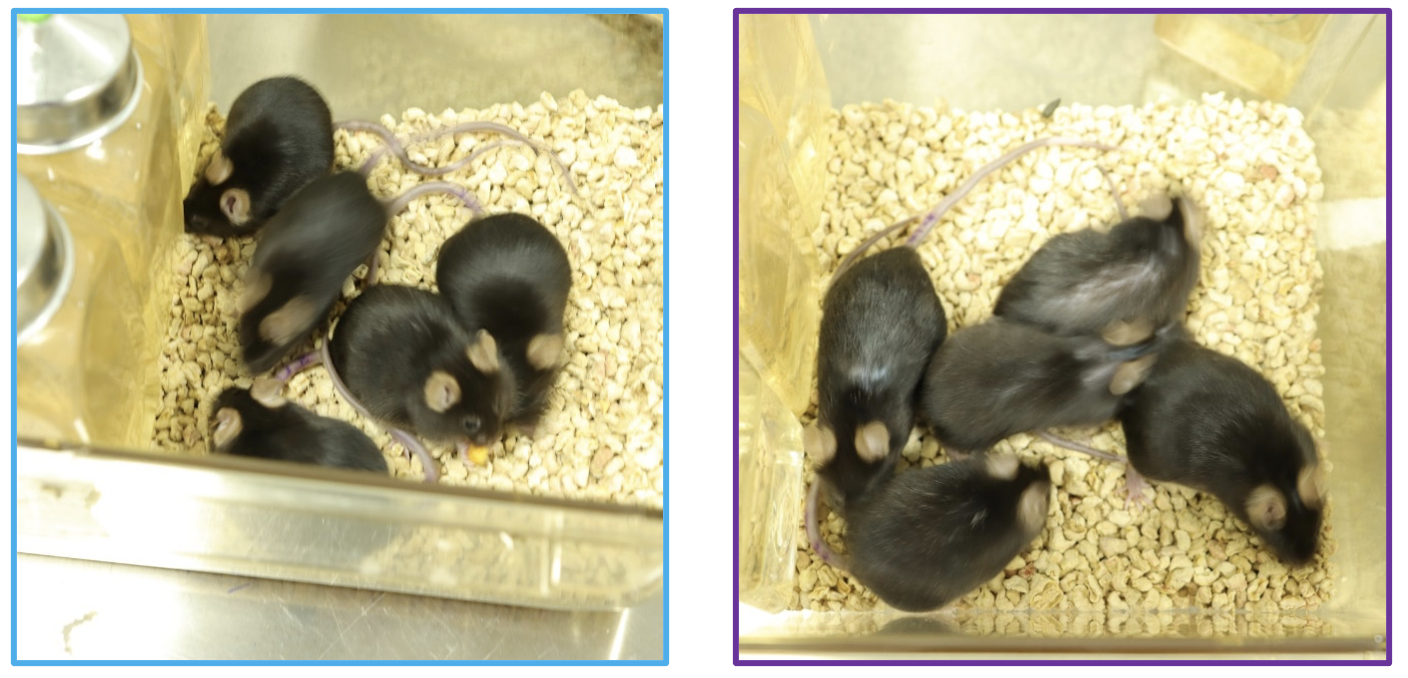
T-168 reduced frailty in aged wildtype mice
- Young mice — control (dashed line)
- Young mice — T-168 (dashed line)
- Old mice — control
- Old mice — T-168
Week
Mechanism of action
T-168 upregulates PGC-1α to restore cellular health
In dopaminergic neurons
- Mitochondrial biogenesis: NRF1, TFAM and mitochondrial mass increase, restoring ATP production
- Mitophagy restored via PINK1/Parkin-mediated clearance of damaged mitochondria
- MFN1-driven mitochondrial fusion
- Lysosomal function & autophagy: TFEB nuclear translocation and restored acidification via V-ATPase assembly
In microglia
- Metabolic switch from glycolysis toward oxidative phosphorylation
- Reduced inflammatory cytokine release (TNF, IL-6)
- More homeostatic phagocytosis — effect disappears when PGC-1α is deleted specifically in microglia
- Enhanced autophagy and clearance of damaged proteins
Clinical
SLEIPNIR: a biomarker-driven platform trial in Parkinson's disease
Biomarkers
- Brain energetics and glucose metabolism (PET and MRI)
- Markers of mitochondrial and lysosomal health (CSF and plasma)
- Markers of disease and neuronal injury (α-syn, NfL; CSF and plasma)
- Markers of inflammation (CSF and plasma)
- CNS penetration
Clinical assessment
- MDS-UPDRS
- Grip strength (handheld dynamometer)
- Axivity AX6 wearable activity tracking
- B-SIT smell test
The biomarker-rich design is intended to establish the relationship between drug exposure, CNS biological activity and clinical outcomes directly in Parkinson's disease patients.
Indication expansion
One upstream mechanism, a broad therapeutic opportunity
Indication opportunities in neurodegeneration
Indication opportunities include orphan and non-orphan diseases including:
Orphan: Amyotrophic Lateral Sclerosis (ALS), Frontotemporal Dementia (FTD), Friedreich's Ataxia (FA), Huntington's Disease (HD)
Prevalent: Alzheimer's Disease (AD), Parkinson's Disease (PD)
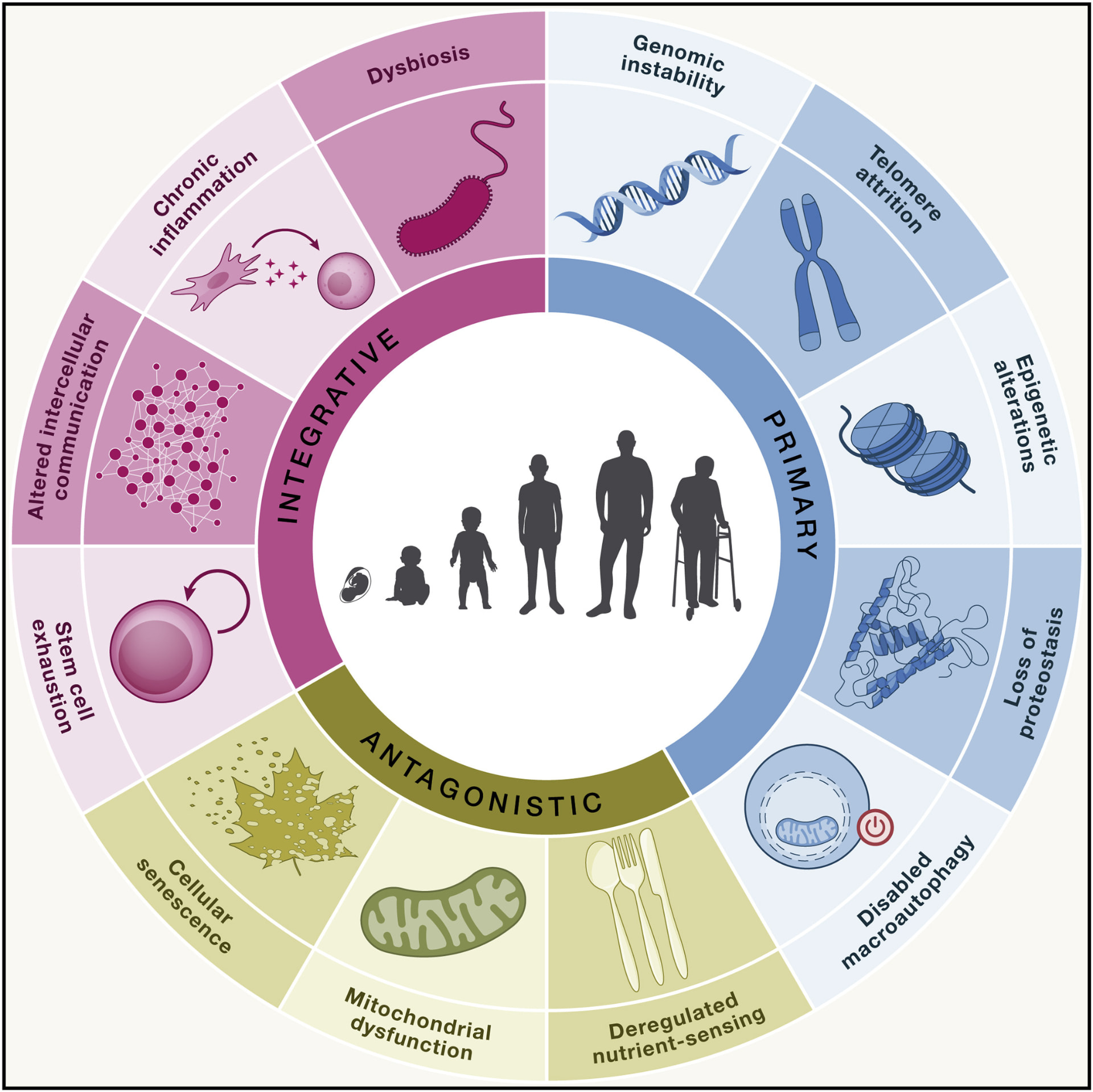
Emerging opportunities: aging, frailty, metabolic and muscle diseases
In aged wild-type mice, T-168 treatment resulted in a 51% lower frailty score, 28% reduction of fat mass with no loss of muscle mass, 25% greater grip strength and 53% greater treadmill endurance versus vehicle-treated aged controls.
Team
Experienced team with a proven track record in drug development
TEAM
 Frederic Godderis, MS
Founder · CEO · Director
 Clarence R. Hurt, PhD
Head of Discovery Research / Medicinal Chemistry
 Andrew Lam, MS
Co-founder · Chief Technology Officer
 Enchi Liu, PhD
Co-founder · Chief Development Officer
 Subhasis Roy, MBA
Chief Financial Officer and Chief Business Officer
Board of Directors
 Taro Inaba, BE, MBA
Managing Partner, Remiges Ventures
 Jackson Streeter, MD
Florida Opportunity Fund / Partner, DeepWork Capital
 Sanjay Kakkar, MD, MPH
Founder · Director · CEO, Tensive SRL · Former Chairman and CEO, Tranquis Therapeutics
- Frederic Godderis, MS
Founder · CEO · Director
Advisors
 Edgar Engleman, MD, PhD
Scientific founder · Professor of Pathology and Medicine, Stanford University · Founder, Vivo Capital
 Charalampos Tzoulis, MD, PhD
Advisor · Professor of Neurology & Neurogenetics, University of Bergen, Norway
Partners
Investors and collaborations

 Managed by






Publications
Selected key literature
(In certain publications, T-168 is also referred to as TQS-168 or ZLN005)
T-168 PHASE 1 PUBLICATION
Hannestad J et al. Clin Transl Sci. 2024;17(11):e70064.
https://doi.org/10.1111/cts.70064
PGC-1α review in neurological diseases
Guo P et al. Regulating PGC-1α: a potential role in neurological disorder and treatment. Eur J Pharmacol. 2025;1001:177750.
https://doi.org/10.1016/j.ejphar.2025.177750
PGC-1 family review
Qian L et al. Peroxisome proliferator-activated receptor gamma coactivator-1 (PGC-1) family in physiological and pathophysiological process and diseases. Signal Transduct Target Ther. 2024;9:50.
https://doi.org/10.1038/s41392-024-01756-w
TFEB in nigral neurons in PD
Moors TE, Morella ML et al. Altered TFEB subcellular localization in nigral neurons of subjects with incidental, sporadic and GBA-related Lewy body diseases. Acta Neuropathol. 2024;147:67.
https://doi.org/10.1007/s00401-024-02707-z
TFEB–ATP6V0C in microglia and in PD
Wang Y, Ma Z et al. Dysregulation of the TFEB-ATP6V0C axis in microglia exacerbates α-synuclein pathology through impaired lysosomal acidification in Parkinson's disease. Cell Death Differ. 2026.
https://doi.org/10.1038/s41418-026-01800-y
Aging hallmarks
López-Otín C, Blasco MA, Partridge L, Serrano M, Kroemer G. Hallmarks of aging: An expanding universe. Cell. 2023;186(2):243-278.
https://doi.org/10.1016/j.cell.2022.11.001
Mitochondria and aging
Weissig V. Mitochondrial dysfunction as the "mother" of all hallmarks of aging. J Mitochondria Plastids Endosymbiosis. 2025;3(1):2560191.
https://doi.org/10.1080/28347056.2025.2560191
T-168 (ZLN) AND PGC-1Α / FERROPTOSIS EFFECTS IN PD
Liu Z et al. LINC-EPS protects against neurodegeneration by driving a PGC-1α-mediated anti-ferroptosis program in Parkinson's disease. Int J Biol Sci. 2026;22(7):3367-3388.
https://doi.org/10.7150/ijbs.128204
T-168 (ZLN) restores dopaminergic neurons in PD
Kaur J et al. ZLN005 Alleviates the Dopaminergic Degeneration in Parkinson's Disease by Upregulating PGC-1α-Dependent Mitochondrial Function. Mol Neurobiol. 2026;63(1):303.
https://doi.org/10.1007/s12035-025-05612-y